UCLA Department of Chemistry and Biochemistry
153AH - Fall 2009 - Instructors: Todd Yeates, Duilio Cascio, Tobias Sayre
Human Transthyretin (TTR) and its association with Thyroxine (T4)
by Naira Barsegyan
|
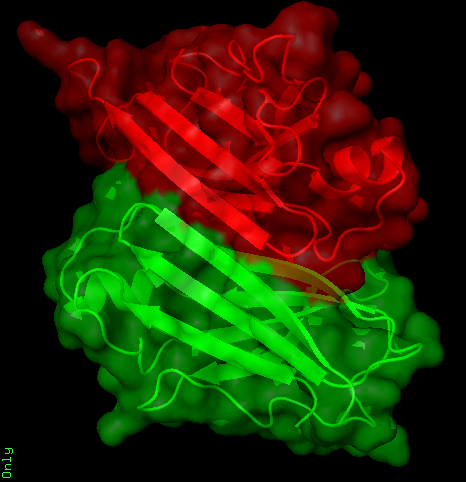
Transthyretin (TTR), also known as prealbumin, is a 127-residue polypeptide rich in β-sheets, though it contains α-helices as well (1,3,5). This overall configuration can be clearly seen in figure 1. It is a transport protein for the hormone thyroxine (T4). It is the primary carrier of T4 in cerebrospinal fluid. TTR not only carries T4, it can also carry retinol (vitamin A) by forming a complex with retinol-binding protein (RBP) (2,5). Various other small molecules have been proven to bind in the T4 binding sites. These include many natural products such as resveratrol, drugs such as diflunisa and flufenamic acid, and toxins such as PolyChlorinated Biphenyls (PCBs). Thyroid hormones are necessary for efficient development and for normal metabolism in mammals. T4 is the main compound produced by thyroid follicles. More than 99% of the circulating hormone is bound to plasma proteins, mainly to T4-binding globulin, TTR, and albumin in humans. TTR is primarily produced in the liver and choroid plexus, which are also the tissues that secrete TTR into the blood and cerebrospinal fluid (4). TTR has been proven to aid in the transfer of the thyroid hormone into tissues, and especially in the transfer of T4 into the brain across the choroid-plexus-cerebrospinal fluid barrier. Yet the exact mechanism in which TTR accomplishes the transfer of T4 has yet to be discovered. TTR is a homotetramer often described as a dimer of dimers. Two monomers interact with one another in order to form a dimer structure that contains an extended beta sandwich. Another identical pair of monomers interacts with the original pair of monomers in order to produce the native homotetrameric structure. Two monomers, or one of the dimers, interact with another dimer to form the homotetrameric structure, as seen in Figure 2. The two identical sets of dimers align opposite to one another and in between the interior of the complex of the two identical sets of dimmers is where the two T4 ligands bind. Figure 3 outlines the surface structure of TTR, along with its active site. Residue methionine 119 is considered the active site for the protein structure. This active site is the location at which the T4 binds. There are however some variants, such as the methionine 30 residue and the asparagine 90 residue (6). Numerous amyloidogenic mutations of the TTR have been discovered. Recent biochemical and pathological studies have shown that the instability of the homoterameric form of TTR can lead to amyloid formation in the tissues of senile systemic amyloidosis (SSA) and familial amyloidotic polyneuropathy (FAP). This is usually due to the presence of a mutation in the TTR and errors in post-translational modifications (1-4). There are also many mutations that are not amyloidogenic but are directly tied to hyperthyroxinemias. Further research is needed to understand how the various mutations in TTR lead to disease. References 1) Saraiva MJ. (1995).Transthyretin mutations in health and disease. Hum. Mutat. 5(3):191-6. 2) Raghu P, Sivakumar B. (2004). Interactions amongst plasma retinol-binding protein, transthyretin and their ligands: implications in vitamin A homeostasis and transthyretin amyloidosis. Biochim Biophys Acta. 1703(1):1-9. 3) Amiram Raz and DeWitt S. Goodman. (1969). The Interaction of Thyroxine with Human Plasma Prealbumin and with the Prealbumin-Retinol-binding Protein Complex. The Journal of Biological Chemistry, 244, 3230-3237. 4) G. Schreiber, A. R. Aldred, A. Jaworowski, C. Nilsson, M. G. Achen and M. B. Segal. (1990). Thyroxine transport from blood to brain via transthyretin synthesis in choroid plexus. J Physiol Regul Integr Comp Physiol. 258: R338-R345. 5) Walter de Gruyter. (2002). Transthyretin as a thyroid hormone carrier: Function revisited. International Congress on Transthyretin (TTR) in Health and Disease. 40(12), pp. 1292-1300. 6) Alves IL, Divino CM, Schussler GC, Altland K, Almeida MR, Palha JA, Coelho T, Costa PP, Saraiva MJ. (1993). Thyroxine binding in a TTR Met 119 kindred. J Clin Endocrinol Metab. 77(2):484-8. |
|


